Free Dicom Viewer Radiology
Posted : admin On 20.12.2020MicroDicom DICOM viewer
- Free Dicom Viewer Radiology
- Free Dicom Viewer Radiology Assistant
- Free Dicom Viewer Radiology Technologists
Latest version
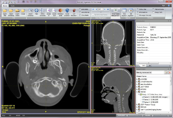
Oct 24, 2019 If you can already view DICOM images within your EHR, you likely won’t require a standalone viewer. However, if your system doesn’t support DICOM, you’re having difficulty communicating with a PACS or RIS system, or if you don’t have PACS/RIS access — or even EHR at all — a free DICOM viewer will help you get started viewing images. DICOM viewing software allows radiology trainees and consultants to view and manipulate medical images (such as radiographs or MRI scans) on their own PC, laptop or tablet. In the hospital environment, this forms part of the picture archiving and communication system (PACS), which doctors will be familiar with! Installation package: MicroDicom DICOM viewer 3.8.1 x86 (4.17 MB 2020-11-25) MicroDicom DICOM viewer 3.8.1 x64 (4.78 MB 2020-11-25). Portable zip package(no installation required).
Installation package:
MicroDicom DICOM viewer 3.8.1 x86 (4.17 MB 2020-11-25)
MicroDicom DICOM viewer 3.8.1 x64 (4.78 MB 2020-11-25)
Portable zip package(no installation required):
MicroDicom DICOM viewer 3.8.1 x86 zip (4.91 MB 2020-11-25)
MicroDicom DICOM viewer 3.8.1 x64 zip (5.80 MB 2020-11-25)
Autorun package for CD/DVD/USB:
MicroDicom DICOM viewer CD/DVD 3.8.1 (10.6 MB 2020-11-25)
MicroDicom Shell Extension
Latest version
Universal installation package for x86 and x64:
MicroDicom Shell extension 3.0.0 (2.49 MB 2020-06-07)
Sample DICOM images
You can download some sample DICOM images from here.
DICOM sample images were temporarily removed.
MicroDicom DICOM viewer older versions:
/bcc-7-for-sony-vegas-crack.html. You can download older versions of MicroDicom DICOM viewer here
View Your Own Medical Scans in 3D
Improve your understanding of your own CT, MRI & PET scans by rendering them in 3D rather than 2D slices in 90 seconds or less!
Your medical images are typically saved in a DICOM format which is coloured in greyscale based on the density of your anatomy.
We have made it easy for you to look at certain anatomy by creating an intuitive slider that allows you to very quickly “strip” away different tissue densities to expose particular areas of interest.
You can view your internal structures such as cartilage, muscle, organs and ultimately your own skeletal system.
Visualise Your 2D DICOM Medical Images In 3D
With Volume Rendered Models
Learn more about your own anatomy with our suite of intuitive DICOM viewers for Desktop on Windows & MacOS
Immersive 3D Visualisation
Using our proprietary volumetric rendering platform, 3Dicom Viewer allows for the rapid conversion of 2D Dicom files into 3D fully immersive volumetric models.
Open Files From Dropbox
Load your DICOM (.DCM) or NiFTI (.nii) directly from your computer or transfer them via Dropbox to your devices to view your own scans rendered locally.
Private & Secure
We protect your personal medical data with 100% offline conversion and viewing, no internet connection or file transfer required. We don’t collect any of your scan data.
Easily View Different Organs
View your scan from any angle and viewpoint, with the ability to render the skin, soft tissue and major organs through to the skeleton.
No Account or Card Required
After signing up to download 3Dicom, we don’t require any account or card details to use the 3Dicom Viewer software.
Free Dicom Viewer Radiology
Developed For Cross Platform
We realise that you want to be able to review your scans whilst on the go and so we’ve included all Windows features for MacOS too.
3Dicom Viewer
How The Software Works
Extensive Viewing Optimisation Tools
Extract the most amount of detail from your scans by adjusting the brightness, contrast, and/or opacity.
Adjusting these values helps to maximise the contrast between surrounding tissue, different anatomical structures and to eliminate ‘noise’ picked up by the CT or MRI scanner during the capture of your medical images.
Reducing the opacity in 3Dicom Viewer creates a X-Ray / Wireframe effect which results in being able to see the location of organs in relation to the skin & external areas.
Free Dicom Viewer Radiology Assistant
Rapid, Offline Volumetric Rendering
Our lightweight 3D viewer isn’t intended as a full clinical DICOM viewer, rather as a fast and easy way to review your 2D analysis in the 3D space. Your time is precious and despite using your own computer’s processing power to render the dicom (.dcm) or NiFti (.nii) files into a 3D volumetric render, we’ve optimised our conversion and rendering process to take less than 60 seconds.
In fact, most scans load in less than 30 seconds, giving you a quick-fire tool to create, review and capture (via screenshot or screen recording) a better understanding of complex cases.
Works with 95% of CT/MRI & PET Scans
We’ve developed our software from the ground up to work entirely offline, protecting your private medical data and allowing remote and rural usage.
This does however mean that we can’t easily draw upon external resources and code libraries to convert all DICOM files. Our team has worked incredibly hard to ensure that we have multiple redundant ways in which to interpret the meta-data in your DICOM scans and currently can convert more than 95% of CT/MRI & PET Scans.
Immersive Movement & Zoom Features
Free Dicom Viewer Radiology Technologists
3Dicom Viewer allows you to get up close and personal with your own anatomy. Using industry standard fluid panning, zooming and rotation tools, you can view the 3D model from any angle.
The converted 3D model, once adjusted for brightness, contrast and the required Hounsfield values, can be ‘entered’ to travel through empty spaces (such as airways and lungs) or inside the soft tissue when it has been ‘stripped’ away to view the skeletal system and major organ systems.
Our Mission To Improve Patient Education
We are committed to developing technology that provides patients and practitioners alike with access to personalised, enhanced medical data to inform better health decisions.
A 2012 study by Mitchell SE, Sadikova E, Jack BW & Paasche-Orlow found that that patients with low health literacy are 1.46 times more likely than patients with adequate health literacy to return to the hospital or emergency department within 30 days.
Providing an easier, more intuitive way in which to perceive radiological data and associated health issues (pathologies) is vital to increasing health literacy, reducing hospital readmissions and empowering patients to make better informed medical decisions.